Our Simple Guide to Ventricular Septal Defects (VSD) in Children
A Ventricular Septal Defect is a hole in the wall of the heart that separates the lower chambers - learn about this issue, how it’s treated & how we help.
A Ventricular Septal Defect is a hole in the wall of the heart that separates the lower chambers. When the hole is large enough, the amount of blood leaking between the chambers can cause permanent damage to the heart and lungs.

What is a Ventricular Septal Defect (VSD)?
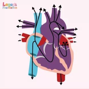
A Ventricular Septal Defect (VSD) is a birth defect of the heart, in which there is a hole in the septum that separates the two lower chambers of the heart.
The hole allows oxygen-rich blood to move back into the lungs instead of being pumped around the body. The mixture of oxygen-rich blood with oxygen-poor blood can increase the blood pressure in the lungs and cause the heart to work much harder to pump the blood.
Is a Ventricular Septal Defect a serious heart condition?
Although a Ventricular Septal Defect is a common congenital heart defect, If left untreated, it can become an extremely serious heart condition. It can increase the risk of other complications such as heart failure, stroke, high blood pressure in the lungs and arrhythmia.
If the hole is small, it may not be detected for a number of years and may only be discovered in adulthood. VSD is rare amongst adults, but it can cause serious complications of the heart.
What is the most common Ventricular Septal Defect in children?
There are four main types of Ventricular Septal Defect, which differ in their location and the structure of the hole(s). These include:
- Membranous – This is the most common type of VSD, making up 80% of cases. These VSDs occur in the upper section of the wall between the ventricles.
- Muscular – These account for about 20% of VSDs in infants, and there is often more than the hole that’s part of the defect.
- Inlet – This is a hole that happens just below the tricuspid valve in the right ventricle and the mitral valve in the right ventricle. This means when blood enters the ventricle, it must pass through the hole connected to the two chambers.
- Outlet (Conoventricular) – A hole is created just before the pulmonary valve in the right ventricle and just before the aortic valve in the left ventricle. This means blood has to go past the hole on its way through both valves.
How common are Ventricular Septal Defects?
Ventricular Septal Defects are the most common type of congenital heart defect in the UK, with around 20% of babies each year being born diagnosed with one.
Ventricular Septal Defects can also occur with an Atrial Septal Defect too, though this is uncommon.
What causes Ventricular Septal Defects?
The causes of Ventricular Septal Defects in some children is still unknown; however, there are various reasons why some babies may be born with the defects. Some babies will develop heart defects because of change in their genes or chromosomes.
Heart defects are also thought to be caused by a combination of other risk factors too, such as things the mother comes into contact with during pregnancy, as well as any medicines she may consume.
How is a Ventricular Septal Defect diagnosed?
A Ventricular Septal Defect is usually diagnosed after a baby is born. However, the size of the hole will usually influence what symptoms are present and if a doctor hears a murmur during a physical examination.
During a physical examination, the doctor may hear a whooshing noise. This will be the murmur. If this is heard, the doctor can ask for more tests such as an echocardiogram to be completed. This is an ultrasound of the heart that will show any problems in relation to its structure.
What are the symptoms of a Ventricular Septal Defect?
If the baby has a large hole in its heart, common symptoms may arise such as:
- Shortness of breath
- Fast or heavy breathing
- Sweating
- Tiredness when feeding
- Poor weight gain
Can a Ventricular Septal Defect be treated?
Treatments for a Ventricular Septal Defect can depend on the size of the hole and the potential problems that it may cause. For many, the hole will close on its own and will not cause any symptoms. If your doctor believes this to be the case, your child will be required to have regular check ups to ensure the hole is closing.
If the hole is slightly larger, your doctor may recommend surgery to close the hole. This can either be open-heart surgery or through cardiac catheterization. After surgery, your child will need to attend regular follow-up appointments to ensure the hole remains closed.
Some children will need medicine to help strengthen their heart muscles, lower the blood pressure and get rid of any excess fluid.
What are the complications of a Ventricular Septal Defect?
Where small Ventricular Septal Defects may not cause any complications, some larger ones can if they’re left untreated. Listed below are some of the complications that could occur.
- Heart failure – With medium and large VSDs, the heart has to work harder to pump the blood to the lungs. Without treatment, heart failure can develop.
- Eisenmenger syndrome – Unrepaired holes in the heart can lead to this complication after many years. Irregular blood flow causes the blood vessels in the lungs to become narrow and stiff. This syndrome permanently damages the blood vessels.
- Endocarditis – This is a rare condition connected to VSD, in which an infection causes life-threatening inflammation of the inner lining of the heart’s chambers and valves.
How long can a child live with a Ventricular Septal Defect?
Children who have been treated for their Ventricular Septal Defect, whether that is through surgery or medicine, are likely to have long life expectancies, usually matching that of someone who has not suffered from a VSD.
For those who have not had it repaired, around 87% will live to around 25 years after their diagnosis. However, this can vary depending on the size and location of the VSD.
How can Lagan’s Foundation help?
At Lagan’s Foundation, we offer respite care to families with Complex Health Needs, specialising in Heart Defects and Feeding Issues. Our respite consists of highly trained staff attending the family’s home and taking care of the child whilst their parents/guardians take a break.
Our experienced carer will take charge of all medications given to the child during their respite hours, as well as any feeding support they may need.
All our staff are specially trained by our charity’s CEO, Training Lead and RCN Clinical Trainer and Assessor, to support children in the North West who have Heart Defects as well as other Complex Health Needs, and during their multiple training days they cover Ventricular Septal Defects specifically in depth.

![Understanding Total Anomalous Pulmonary Venous Return [TAPVR]](/wp-content/uploads/2025/04/Understanding-Total-Anomalous-Pulmonary-Venous-Return-FI-312x312-c-default.jpg)


