
What is Tricuspid Atresia?
Tricuspid Atresia, a condition present at birth, is a heart defect in which your tricuspid valve does not exist. Without this valve, blood can’t flow through the heart and body normally.
Multiple surgeries are required to treat Tricuspid Atresia and thanks to modern medicine, many babies who are treated for the defect will go on to live healthy, adult lives.
Understanding Tricuspid Atresia in children
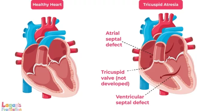
Tricuspid Atresia is a congenital heart defect where the tricuspid valve is either underdeveloped or absent, leading to a small right ventricle and restricted blood flow to the lungs.
In a normal heart, the tricuspid valve lets blood flow from the right atrium into the right ventricle. When the right ventricle pumps blood into the lungs, the tricuspid valve closes. This prevents blood from flowing back into the right atrium.
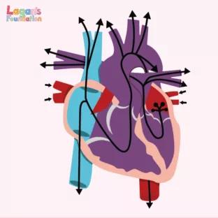
The right side of the heart gets oxygen-poor blood from the body and pumps it to the lungs, which then add fresh oxygen to the blood. The oxygen-rich blood then returns to the left side of the heart which is then pumped out to the rest of the body.
In a heart with Tricuspid Atresia, solid tissue sits between the right atrium and right ventricle. Since the blood in the right atrium cannot move through the tricuspid valve, the wall separating the right and left sides of the heart don’t form properly.
Many babies born with Tricuspid Atresia will also be born with:
- Atrial Septal Defect – A hole between the right and left atrium
- Ventricular Septal Defect – A hole between the right and left ventricles
The signs and symptoms Tricuspid Atresia
Soon after birth, a baby born with Tricuspid Atresia may show a variety of signs and symptoms.
These symptoms can include:
- Bluish skin, known as cyanosis
- Fast and hard breathing
- Difficulty feeding
- Tiring easily when feeding
- Being less active than most babies
These symptoms are also often associated with other congenital heart defects too, which is why an early diagnosis is so important.
How is Tricuspid Atresia diagnosed?
Tricuspid Atresia is sometimes diagnosed through an ultrasound scan before birth. Foetal echocardiograms are also sometimes used as well to give midwives more information and help them to plan immediate treatment.
A pulse oximeter test will usually be conducted after the baby is born by using a light on a fingertip or toe. This test will show that the baby’s blood is not carrying as much oxygen as expected.
Other tests used can include:
- X-rays
- Electrocardiogram
- Echocardiogram
The treatment for Tricuspid Atresia
Treatment for Tricuspid Atresia will usually combine medicine, surgery and cardiac catheterisation to improve the blood flow to the lungs.
Surgery for Tricuspid Atresia is carried out in stages as your child grows. The type and timing of the surgery will depend on the circumstances of your child and which other heart defects they may have. Some children may not have any surgery until they are a few years old, but most will need some form of surgery soon after birth.
These surgical steps, also known as the single ventricle pathway, are as follows:
- Aged 2 weeks old or less – A Blalock-Taussig-Thomas (BTT) shunt is used to redirect some of the blood from the left ventricle from the body to the lungs. Alternatively, if the blood flow to the lungs is too high, a band is placed around the pulmonary artery to lessen the blood flow and prevent lung damage.
- Aged 4-6 months old – The next procedure is known as the Glenn Procedure and allows blood returning from the upper part of the body to flow directly into the lungs. The BTT shunt is removed at the same time.
- Aged 1.5-3 years – The Fontan Procedure channels blood from the lower half of the body to the lungs so the heart pumps oxygen-rich blood to the body only. Blood returning from the body flows to the lungs before passing through the heart.
Based on your child’s diagnosis and if they have any other heart conditions, doctors will decide which surgery is most appropriate and at what age.
Cardiac catheterisation can also be used to make or enlarge openings in the hole between the two atria and between the two ventricles.
Medicine, called Prostaglandin E1, may also be used to keep the ductus arteriosus open. This is a normal blood vessel that connects the aorta and pulmonary artery, the two major arteries that carry blood away from the heart.
What happens as your child grows up with Tricuspid Atresia
As we’ve mentioned, children may need regular surgery as they grow in order to improve blood flow and oxygen levels. Even children who have received surgery at a young age will require lifelong monitoring to ensure their heart is working as it should be.
However, with the correct medical care and follow-ups, children with Tricuspid Atresia can go on to live long, healthy lives.
Home monitoring can help to give your child better outcomes at life and between surgeries. Regular monitoring you can do yourself include:
- Weighing your child regularly
- Learning about your child’s condition
- Keeping track of your child’s eating
- Communicating with healthcare providers
- Administering at home medications
Explore Related:
![Understanding Total Anomalous Pulmonary Venous Return [TAPVR]](/wp-content/uploads/2025/04/Understanding-Total-Anomalous-Pulmonary-Venous-Return-FI-312x312-c-default.jpg)
Lagan’s Cycle Challenge is back!
Join us for the landmark 10th year of Lagan’s Cycle Challenge – an event that’s bigger and better than ever!
Read more
A Simple Guide to Transposition of the Great…
Different types of Transposition of the Great Arteries
Read more